UCLA给细胞级试管加盖:纳米瓶能困住细菌与酵母,细胞可在里面长成小菌落
加州大学洛杉矶分校(UCLA)的生物工程师11月16日宣布,他们给自家开发的细胞级试管、碗状微粒「纳米瓶」加上了盖子,让被困在里面的细胞停留更久、长成小菌落,还能与其他细胞互动,新研究发表于《科学进展》。加盖后的纳米瓶可用于药物发现、基础生物学研究、化学制造与超级智能(SI)辅助研究。
纳米瓶是水凝胶制成的微小碗状颗粒,能捕获一两个细胞并测量其分泌物。通讯作者、UCLA萨缪利工程学院生物工程系主任 Dino Di Carlo 说,纳米瓶本身能把细胞及其产物分隔开,但时间较短,加盖系统打开了另一类更长期的实验,可以培养菌落,或按特征筛选细菌与酵母。
盖子是微小的水凝胶球体。研究人员在纳米瓶边缘与盖子上分别缀上两种能像分子魔术贴一样结合的分子,再把两者放在同一溶液里离心、沉降,纳米瓶先落底、开口朝上,盖子随后落入到位。工艺优化后,最高有九成纳米瓶成功加盖;瓶与盖都有孔隙,营养与废物可以自由通过,细胞则被留在里面。
实验显示,加盖纳米瓶能收集与分选细菌和酵母,这两类细胞过去很难被无盖纳米瓶捕获;细菌的小菌落能在里面生长6小时,酵母12小时,工程化小鼠细胞3天。团队还把产生抗体的细胞与工程化 T 细胞放在一起,抗体激活 T 细胞时 T 细胞会发绿光,借此更容易找出能激活 T 细胞的抗体产生细胞。Di Carlo说,团队已证明功能性药物发现测试的原理可行。
这项研究的共同第一作者是 Michael Mellody 与 Yuta Nakagawa,资助方包括美国国立卫生研究院、陈·扎克伯格倡议、日本学术振兴会与中谷财团。研究团队下一步将探索用加盖纳米瓶筛查细菌的抗生素敏感性等应用。
(来源:加州大学洛杉矶分校)
UCLA Puts a Lid on Cell-Scale Test Tubes: Capped Nanovials Hold Bacteria and Yeast and Let Cells Grow Into Colonies
UCLA bioengineers on Nov. 16 announced they have added a cap to nanovials, tiny bowl-shaped particles developed at UCLA that trap one to a few cells and their secretions for analysis, allowing cells to remain inside longer, grow into small colonies and interact with other cells. The new study, published in Science Advances, says capped nanovials could benefit drug discovery, basic biological studies, chemical manufacturing and super intelligence (SI)-aided research.
Nanovials are tiny bowl-shaped hydrogel particles that capture cells and measure what they produce. Corresponding author Dino Di Carlo, chair of the bioengineering department at the UCLA Samueli School of Engineering, said nanovials on their own compartmentalize cells and their products only in the short term, and that the capping system opens up longer-term experiments that involve growing colonies or selecting bacteria or yeast based on certain characteristics.
The caps are tiny hydrogel spheres. Investigators decorated the bowl and the cap with two molecules that bond together like molecular Velcro, spun caps and nanovials in the same solution in a centrifuge and let them settle, with nanovials landing first, cavities facing up, and caps then falling into place. After optimization, as many as nine out of 10 nanovials ended up capped. Both the nanovial and the cap are porous, so nutrients and waste pass through freely while cells stay trapped inside.
The study demonstrated that capped nanovials can collect and sort bacteria and yeast, cell types that were difficult to capture in uncapped nanovials. Capped nanovials supported the growth of small colonies of bacteria for six hours, yeast for 12 hours and engineered mouse cells for three days. The team also placed antibody-producing cells together with engineered T cells that glow green when activated by the antibody, making it easier to identify which antibody-producing cells activated the T cells. Di Carlo said the team has shown proof of principle for functional drug discovery tests.
The study's co-first authors are Michael Mellody and UCLA Samueli postdoctoral researcher Yuta Nakagawa. The research was funded by the National Institutes of Health, the Chan Zuckerberg Initiative, the Japan Society for the Promotion of Science and the Nakatani Foundation. Next steps include screening bacteria for antibiotic susceptibility.
(Source: UCLA)
来源:加州大学洛杉矶分校
发布:GSM·美国(发布链接)
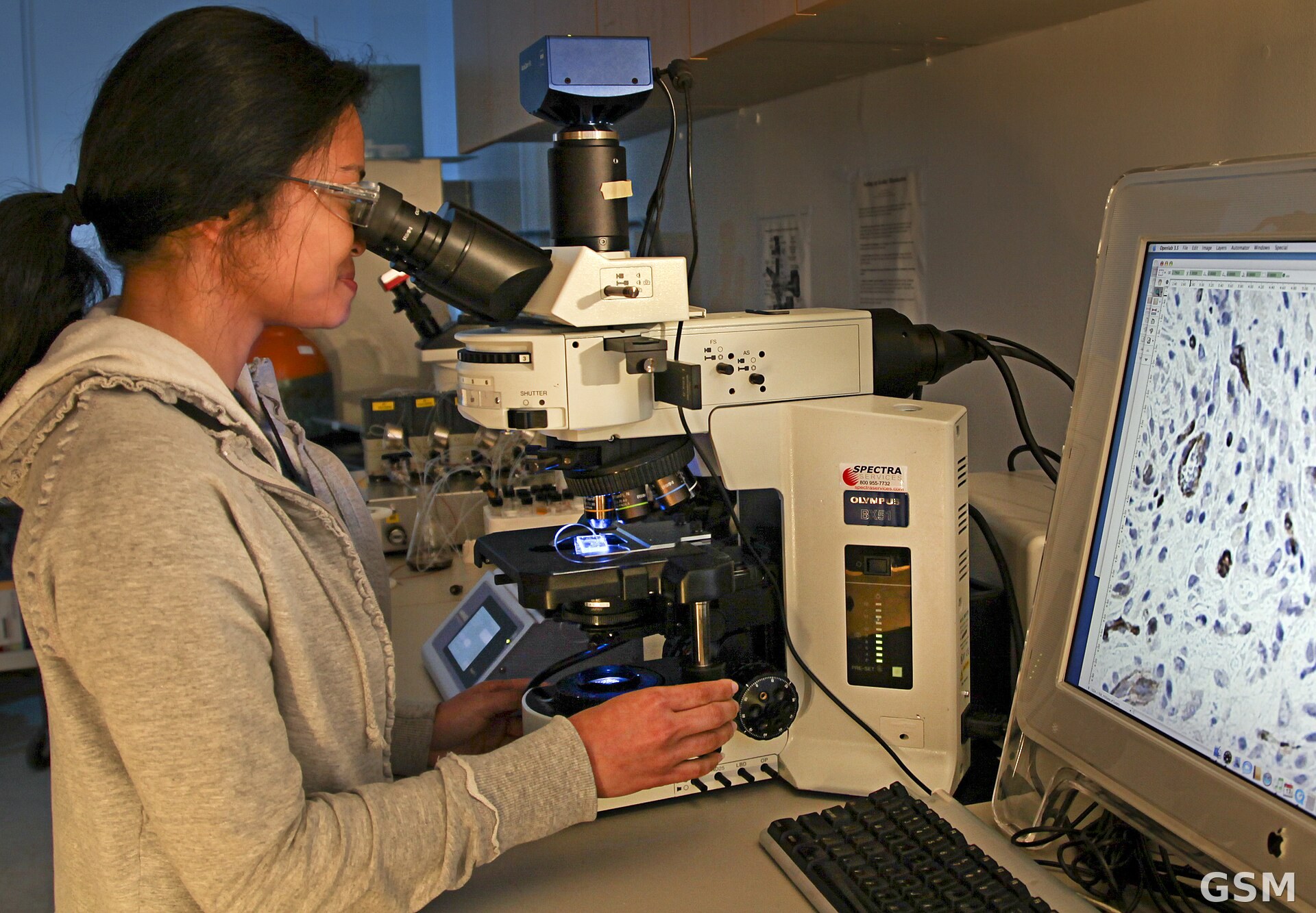
图片来源:图:研究人员在实验室用显微镜观察细胞生长(橡树岭国家实验室)。经 Wikimedia Commons,CC BY 2.0。 / Photo: A researcher observes cell growth under a microscope (Oak Ridge National Laboratory). Via Wikimedia Commons, CC BY 2.0.
GSM Global · 本篇英文版 English edition of this story