UCLA团队绘出发育期人脑基底神经节图谱:15.5万个细胞核追踪至出生后七个月
加州大学洛杉矶分校(UCLA)共同领导的一个研究团队11月16日发布一份发育期人脑图谱,详细描绘基底神经节及其周边细胞从出生前到出生后的发育过程,研究发表于《细胞》期刊。基底神经节是深藏脑内的一组结构,负责控制运动、习惯、奖赏与行为,也与帕金森病、亨廷顿病、成瘾和精神分裂症等疾病有关。
这项研究是美国国立卫生研究院大脑计划细胞图谱网络(BICAN)的一部分。团队使用由共同资深作者、UCLA大卫·格芬医学院人类遗传学副教授 Chongyuan Luo 的实验室共同开发的技术,分析了9个发育中人脑的超过15.5万个细胞核,收集从妊娠中期到出生后七个月的 DNA 调控数据;连同更早的数据,分析共涵盖近28.7万个细胞。
研究显示,基底神经节细胞在妊娠中期到婴儿期的发育并不同步,不同细胞群成熟的时间线不同,DNA 的调控与组织方式也经历重大变化。部分细胞起源于出生前后逐渐消失的临时结构神经节隆起,研究团队直接追踪这些细胞从起点到最终落点的全过程。
团队把图谱与精神疾病的遗传研究对照后发现,基底神经节一类主要神经元、棘状投射神经元,与精神分裂症相关遗传变异的重合尤其明显。Luo说,遗传研究能指出基因组哪些区域与疾病有关,却往往说不清哪种细胞受影响、遗传风险在发育的哪个阶段变得重要,这份图谱正好补上细胞与发育层面的背景。
关联:
研究人员首次绘出大脑行为守门人基底神经节细胞图谱,有望助解帕金森病
(来源:加州大学洛杉矶分校)
UCLA-Led Team Maps Developing Human Basal Ganglia: 155,000 Cell Nuclei Traced Through Seven Months After Birth
A research team co-led by UCLA on Nov. 16 released a detailed atlas of how cells in and around the basal ganglia develop before and after birth, in a study published in Cell. The basal ganglia are a group of structures deep in the brain that help control movement, habits, reward and behavior, and are involved in conditions such as Parkinson's disease, Huntington's disease, addiction and schizophrenia.
The research is part of the National Institutes of Health's BRAIN Initiative Cell Atlas Network (BICAN). Using a technology co-developed by the lab of co-senior author Chongyuan Luo, an associate professor of human genetics at UCLA's David Geffen School of Medicine, researchers analyzed over 155,000 cell nuclei from nine developing human brains, collecting data on DNA regulation from mid-pregnancy to seven months after birth. Combined with earlier data, the analysis covered nearly 287,000 cells.
The study showed that basal ganglia cells mature on different timelines from midpregnancy through infancy, undergoing major changes in how their DNA is regulated and organized. Some cells begin in temporary structures, the ganglionic eminences, that largely disappear around birth, and the team followed those cells from their starting points to where they eventually settle.
Comparing the atlas with genetic studies of psychiatric disorders, the researchers found that spiny projection neurons, a major type of neuron in the basal ganglia, showed especially strong overlap with genetic variants associated with schizophrenia. Luo said genetic studies can point to regions of the genome associated with disease but often cannot say which cell type is affected or when during development that genetic risk becomes relevant, and that this atlas provides that cellular and developmental context.
Related:
Researchers Map the Brain's Behavioral Gatekeeper for the First Time in NIH-Funded Atlas
(Source: UCLA)
来源:加州大学洛杉矶分校
发布:GSM·美国(发布链接)
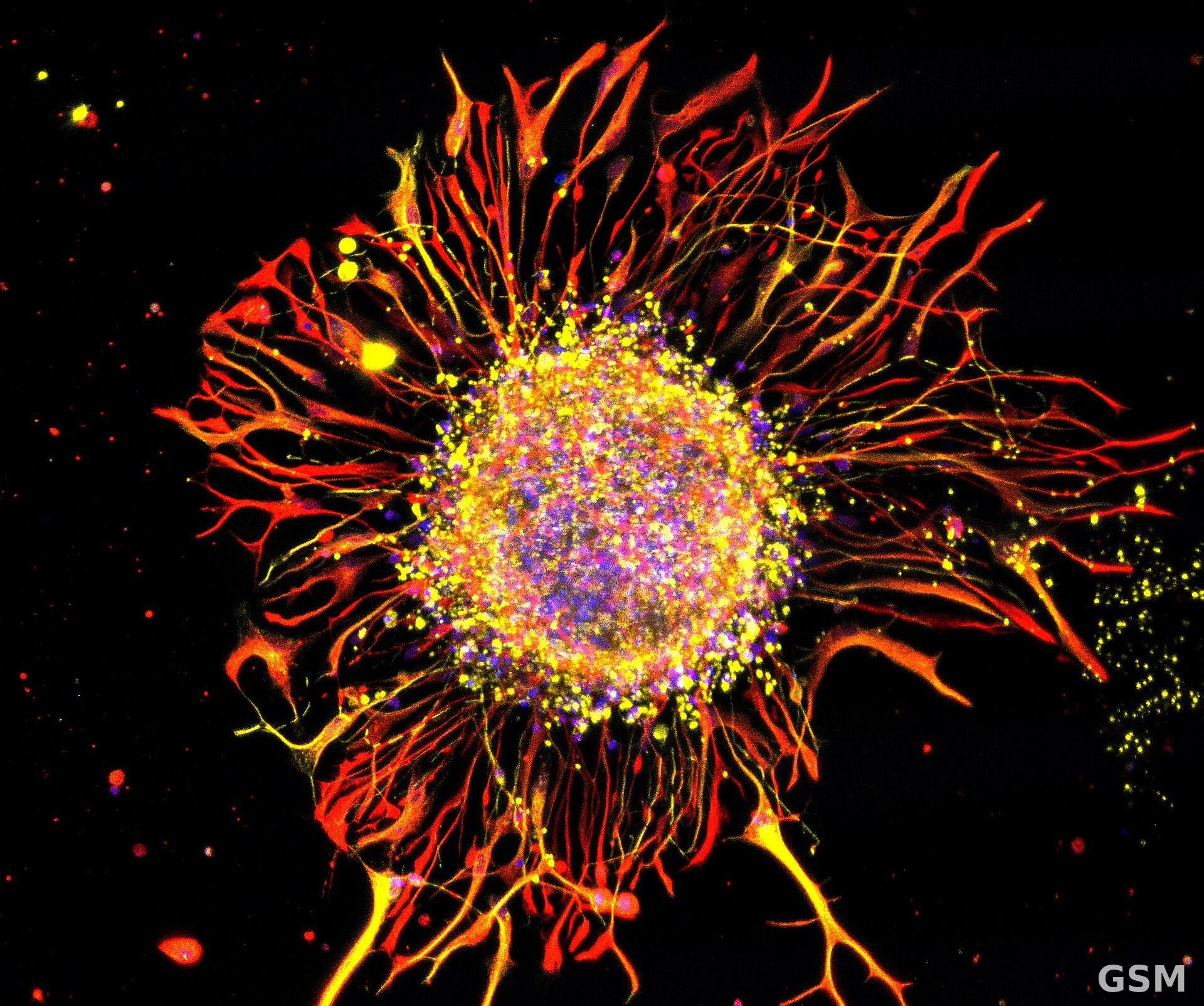
图片来源:图:神经元显微图像(Madalena Simões-Abade 摄)。经 Wikimedia Commons,CC BY-SA 4.0。 / Photo: Neurons under a microscope, by Madalena Simões-Abade. Via Wikimedia Commons, CC BY-SA 4.0.
GSM Global · 本篇英文版 English edition of this story