视网膜神经纤维束高分辨率多深度量化建立规范数据库:18名健康志愿者三项研究、浅层纤维束平均宽12%,《科学报告》11月13日刊出
《科学报告》于 2026 年 11 月 13 日刊出法国研究团队的一项研究,为视网膜神经纤维层建立高分辨率多深度量化方法与规范数据库,论文数字对象标识为 10.1038/s41598-026-78810-w。
视网膜神经纤维层由汇向视神经头的轴突束构成,其厚度是诊断与监测青光眼及神经退行性疾病的关键生物标志物。单根神经纤维束的高分辨率正面成像能给出常规光学相干断层扫描之外的形态信息,但此前缺乏自动分析工具与规范数据。
研究团队开发了图像处理管线,自动量化纤维束的轨迹、弯曲度、方向与宽度,并用自适应光学扫描激光检眼镜与全场光学相干断层扫描,对 18 名健康志愿者建立规范数据库,受试者来自三项研究,各组人数为 15 人、11 人与 11 人。
轨迹分析显示,实测轨迹与现有数学模型存在系统性偏差,团队据此提出两个修正模型。纤维束在较浅的视网膜深度平均宽 12%,且纤维束宽度随年龄显著减小。
论文作者为 Clémentine Callet、Maxime Bertrand、Kate Grieve 等人。研究于 2026 年 7 月 16 日收稿、10 月 28 日接受,处理管线与规范数据意在为后续青光眼与神经退行性疾病的影像研究提供参照。
(来源:《科学报告》)
High-Resolution Multi-Depth Quantification of the Retinal Nerve Fiber Layer Builds a Normative Database: 18 Healthy Volunteers Across Three Studies, With Bundles on Average 12 Percent Wider at Shallower Depths, Published in Scientific Reports on November 13
Scientific Reports published a study on November 13, 2026 presenting high-resolution multi-depth quantification of the retinal nerve fiber layer and a normative database for it, under DOI 10.1038/s41598-026-78810-w. The retinal nerve fiber layer, composed of axon bundles converging toward the optic nerve head, has a thickness that is a key biomarker for diagnosing and monitoring glaucoma and neurodegenerative diseases. High-resolution en face imaging of individual nerve fiber bundles offers morphological information beyond conventional optical coherence tomography, yet automated analysis tools and normative data have been lacking. The researchers developed image processing pipelines for automated quantification of bundle trajectory, tortuosity, orientation and width, and established a normative database from 18 healthy volunteers enrolled across three studies, with group sizes of 15, 11 and 11, imaged using adaptive optics scanning laser ophthalmoscopy and full-field optical coherence tomography. Trajectory analysis revealed systematic deviations from existing mathematical models, leading the team to propose two refined models. Bundles were on average 12 percent wider at shallower retinal depths, and bundle width decreased significantly with age. The paper, by Clémentine Callet, Maxime Bertrand, Kate Grieve and co-authors, was received on July 16, 2026 and accepted on October 28, 2026. The pipelines and normative data are intended as a reference for later imaging research on glaucoma and neurodegenerative diseases.
(Source: Scientific)
来源:《科学报告》
发布:GSM·美国(发布链接)
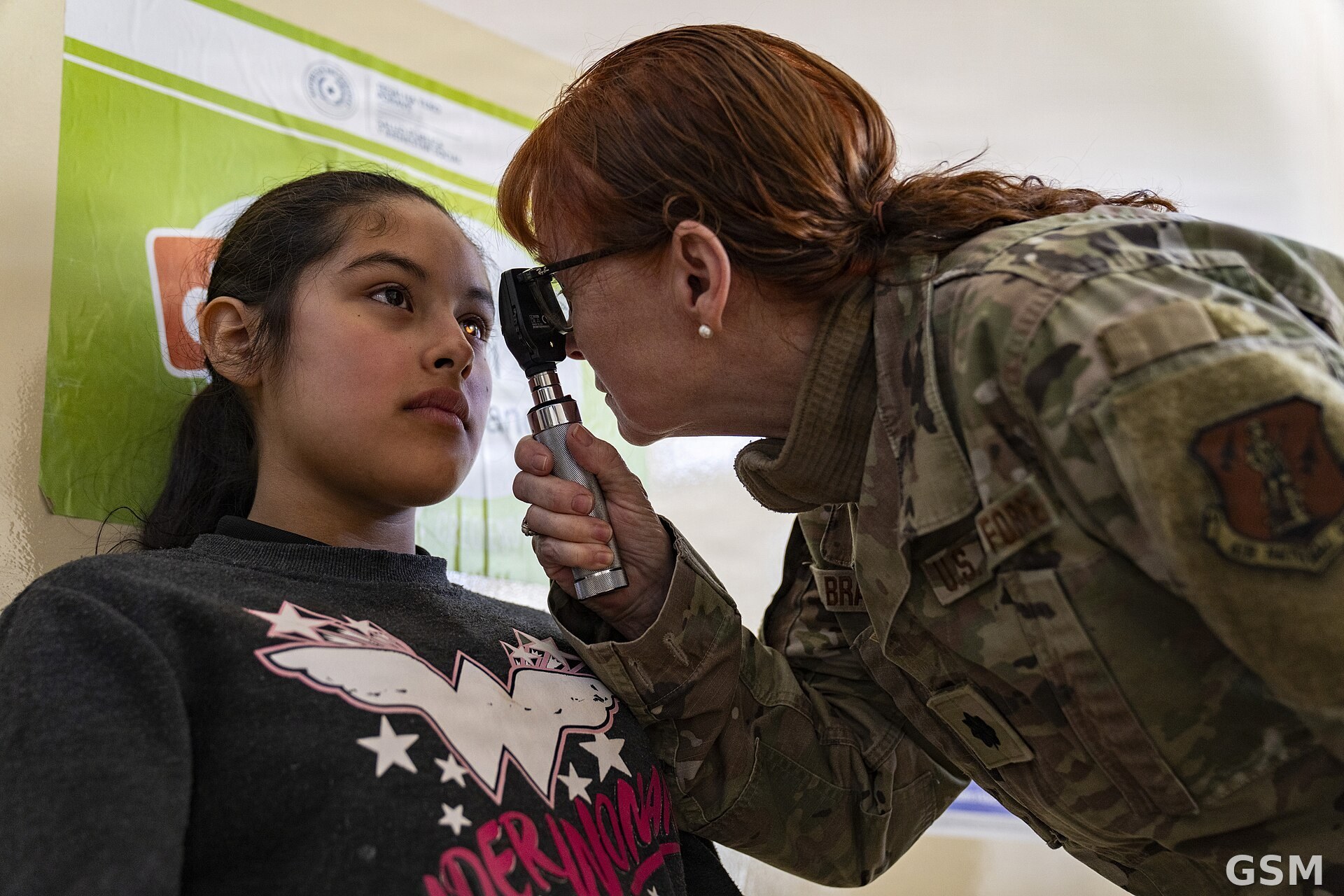
图片来源:图:眼科检查场景。U.S. Air Force photo by Andrea Jenkins,公共领域,经 Wikimedia Commons。 / Photo: U.S. Air Force photo by Andrea Jenkins, public domain, via Wikimedia Commons.
GSM Global · 本篇英文版 English edition of this story