小鼠大脑新图谱11月13日发表:次级运动皮层按身体部位分区,连接初级感觉与视觉皮层
一项11月13日刊于《自然通讯》(Nature Communications)的研究,为小鼠大脑如何表征身体绘制了新的图谱。研究题为《内在神经动力学揭示小鼠次级运动皮层的身体部位特异地形图》,通讯作者团队为 Weihao Xu 等,论文数字对象标识为 10.1038/s41467-026-78900-3。
研究发现,小鼠次级运动皮层(M2)内部存在按身体部位区分的地形组织。研究人员并确认,来自身体部位特异的初级体感皮层(S1)与初级视觉皮层(V1)的结构连接与 M2 的这种地形组织相吻合。
研究团队表示,理解大脑如何协调多个身体部位完成复杂动作,是神经科学的一个基本问题,例如协调手臂、手、头与嘴唇喝一勺汤。这项小鼠研究为回答这一问题提供了新的生物学蓝图。
New mouse brain map published November 13: secondary motor cortex is organised by body part, linked to primary sensory and visual areas
A study published on November 13 in Nature Communications presents a new biological blueprint of how the body of a mouse is represented in its brain. The paper, by Weihao Xu and coauthors, is titled Intrinsic neural dynamics reveal a body-part-specific topography in mouse secondary motor cortex, with DOI 10.1038/s41467-026-78900-3.
The study found a body-part-specific topography inside the mouse secondary motor cortex, known as M2. Structural connectivity from body-part-specific primary somatosensory cortex (S1) and primary visual cortex (V1) to M2 confirmed the M2 topography.
The researchers said a fundamental question in neuroscience is how complex motions that use more than one body part are performed, such as coordinating arm, hand, head and lips to sip a spoonful of soup. The mouse study offers a new map for investigating that question.
来源:自然通讯
发布:GSM·美国(发布链接)
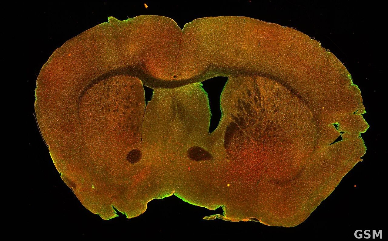
图片来源:图中是小鼠大脑的冠状切片图,相关研究绘制了次级运动皮层的身体部位图谱。Syed07, CC BY 4.0, via Wikimedia Commons
GSM Global · 本篇英文版 English edition of this story