新数学方法让 X 射线三维成像分辨 20 纳米结构:隆德大学团队成果发表于 Optica,分辨率提升约 3 倍
一个国际研究团队开发出一种新方法,可显著提高 X 射线纳米断层成像的三维图像质量。成果由第一作者维克托·尼基京等人发表于 Optica 期刊,隆德大学数学研究员马库斯·卡尔松为末位作者,主导了其中的数学与算法工作。
卡尔松在机构发布中说,这项工作表明,限制因素不只在 X 射线光学器件,也在用于重建图像的数学方法。
新方法可分辨小至 20 纳米的结构。作为对照,人的头发直径约为 8 万至 10 万纳米。在本次研究中,图像分辨率较此前提升约 3 倍,突触、线粒体等亚细胞精细结构得以在三维中显现。
研究者指出,观察纳米级结构通常使用电子显微镜,但样本必须切成极薄的切片才能三维成像,且过程会对材料造成永久损伤。X 射线技术则可对更大的样本做无损三维成像,难点在于从海量测量数据中提取全部信息。
卡尔松说,借助更好的数学模型与优化算法,可以从同类测量数据中提取显著更多的信息,新方法为生物成像及其他需要无损三维研究材料的领域打开了可能性。论文数字对象标识符为 10.1364/optica.603920,方法名称为双线性 Hessian 方法。
New Mathematical Method Sharpens X-Ray 3D Imaging to 20 Nanometers: Lund Team Reports About Threefold Gain in Optica
An international team has developed a method that produces significantly higher quality three-dimensional X-ray images for X-ray nanotomography. The paper by Viktor Nikitin and colleagues appears in Optica, with Marcus Carlsson, a mathematics researcher at Lund University, as last author leading the mathematical and algorithmic work.
Carlsson said the work shows the limitation has not only been in the X-ray optics, but also in the mathematics used to reconstruct the image.
The new method can distinguish structures down to 20 nanometers. By comparison, a human hair is between 80,000 and 100,000 nanometers thick. In the current study, image resolution improved by around threefold, making fine structures such as synapses and mitochondria visible in three dimensions.
Researchers normally use electron microscopy at the nanoscale, but samples must be cut into very thin slices for three-dimensional imaging and the process permanently damages the material, the team noted. X-ray imaging can examine much larger samples in three dimensions without cutting them apart, with the challenge lying in extracting information from enormous data volumes.
Carlsson said better mathematical models and optimization algorithms can extract significantly more information from the same measurement data, opening possibilities in biological imaging and other fields needing nondestructive three-dimensional study. The paper, using the bilinear Hessian method, carries digital object identifier 10.1364/optica.603920.
发布:GSM·美国(发布链接)
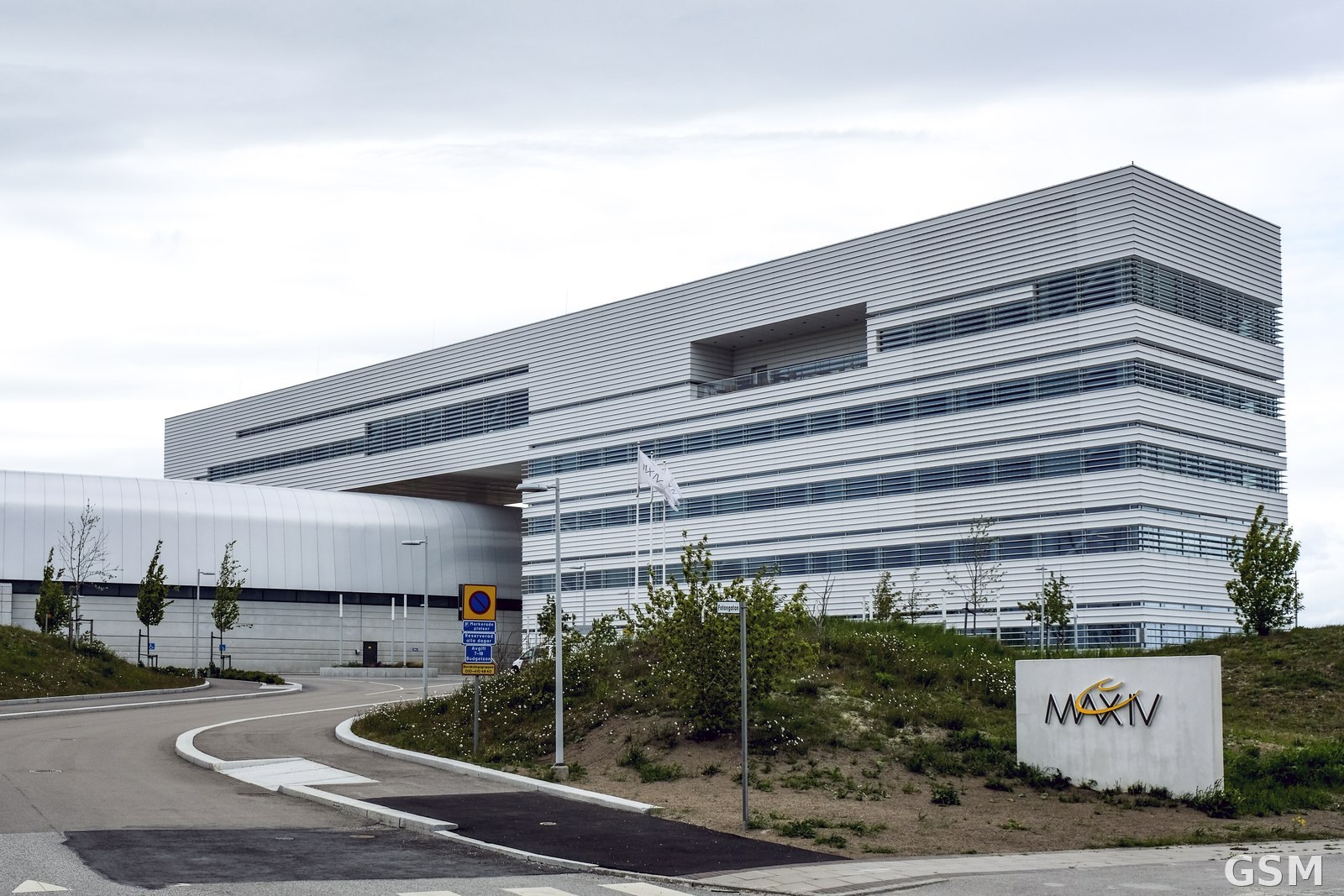
图片来源:图:位于瑞典隆德的 MAX IV 同步辐射实验室建筑。Johan Wessman 摄,CC BY 2.0,经 Wikimedia Commons / Photo: 20200517 Max IV i Lund 002 (49913522522). By Johan Wessman, CC BY 2.0, via Wikimedia Commons
GSM Global · 本篇英文版 English edition of this story