加州大学圣地亚哥分校《Nature Communications》11月16日论文:肺与肠类器官发育中线粒体网络形态与运动特征可预测发育阶段
美国加州大学圣地亚哥分校 Johannes Schöneberg 团队在《Nature Communications》发表论文,11月16日刊出,通讯作者为 Schöneberg,第一作者包括 Gillian McMahon 与 Dhruv Agarwal 等,作者单位为该校药理学系与生物化学和分子生物物理学系。
线粒体在细胞内形成动态的三维网络,其形态与运动随细胞状态、类型与命运而变化,但单个状态的表型此前多是孤立描述,尚不清楚线粒体形态与运动如何贯穿完整的人体组织发育过程变化。团队以人干细胞衍生的分支型肺类器官与肠类器官模拟发育中的人体器官,使这一过程可以在实验室中观察。
团队用晶格光片显微镜在从干细胞到成熟类器官组织的关键发育时间点成像,追踪线粒体网络的三维形态与运动随时间的变化,即所谓四维记录。结果显示,线粒体网络会采取随发育阶段而变的形态与运动特征组合,线粒体特征与细胞特征可以用来预测样本所处的发育阶段。
团队还发现,只做二维测量会掩盖只有在三维加时间的记录中才能分辨的特征;整合后的图谱把线粒体表型贯穿人体组织发育全程描绘出来。研究由美国国立卫生研究院院长新创新者奖(GM150022)与国家生物医学成像和生物工程研究所培训奖(T32EB009380)支持。作者说明,该工作建立的是发育过程的表型图谱与预测关系,不涉及疾病治疗效果。
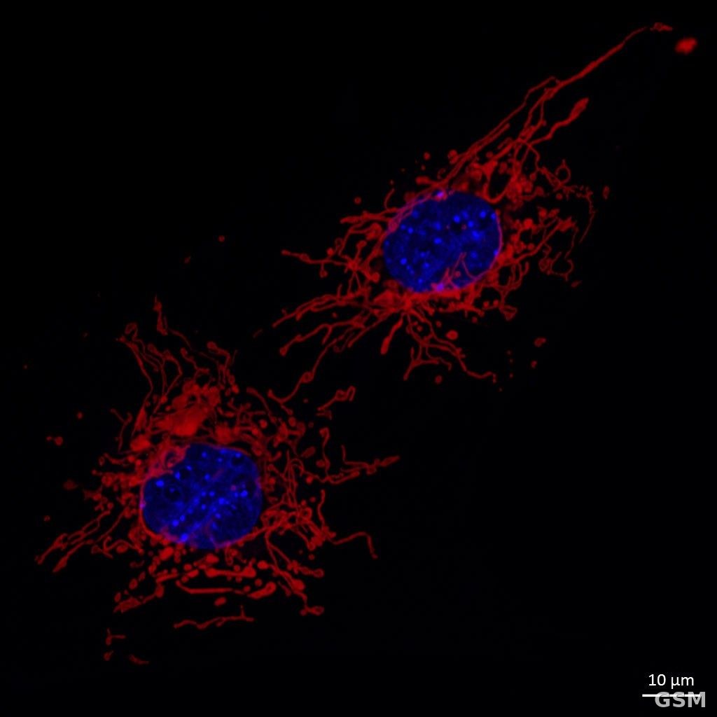
图中是小鼠胚胎成纤维细胞的线粒体(红色)与细胞核(蓝色)荧光显微图,线粒体在细胞内连成的网络正是本研究追踪的对象。
UC San Diego Team in Nature Communications, November 16: Mitochondrial Network Morphology and Motility Predict Developmental Stage Across Human Lung and Intestinal Organoid Development
A team led by Johannes Schöneberg at the University of California, San Diego, in the United States has published a paper in Nature Communications on November 16, with Gillian McMahon and Dhruv Agarwal among the lead authors and affiliations in the university's Department of Pharmacology and its Department of Biochemistry and Molecular Biophysics.
Mitochondria form dynamic three-dimensional networks whose morphology and motility vary with cell state, cell type and cell fate, but phenotypes had previously been described only for isolated states, and how mitochondrial form and movement change across the full course of human tissue development was unclear. The team used human stem cell-derived branching lung and intestinal organoids, which mimic developing human organs, to make the process accessible in the laboratory.
Using lattice light-sheet microscopy, the team imaged the organoids at key developmental time points from stem cell to mature tissue, tracking mitochondrial networks in three dimensions over time. The networks adopted morphology and motility signatures that varied with developmental stage, and mitochondrial and cellular features together could be used to predict which developmental stage a sample was in.
The team also found that two-dimensional measurements obscure features that are resolved only in three dimensions over time, and it assembled the results into an integrated map of mitochondrial phenotypes across human tissue development. The study was supported by an NIH Director's New Innovator Award (GM150022) and an NIH National Institute of Biomedical Imaging and Bioengineering training award (T32EB009380). The authors frame the work as a phenotypic map and a predictive relationship for development, not as a disease treatment finding.
发布:GSM·美国(发布链接)
图片来源:Wikimedia Commons, File:MEF cell mitochondria.jpg — Author: Institute of Molecular Medicine I, University of Düsseldorf. Licensing: CC BY 4.0.
GSM Global · 本篇英文版 English edition of this story