MD安德森绘制0期乳腺癌细胞图谱:逾85万个细胞显示浸润特征出现得比预想更早
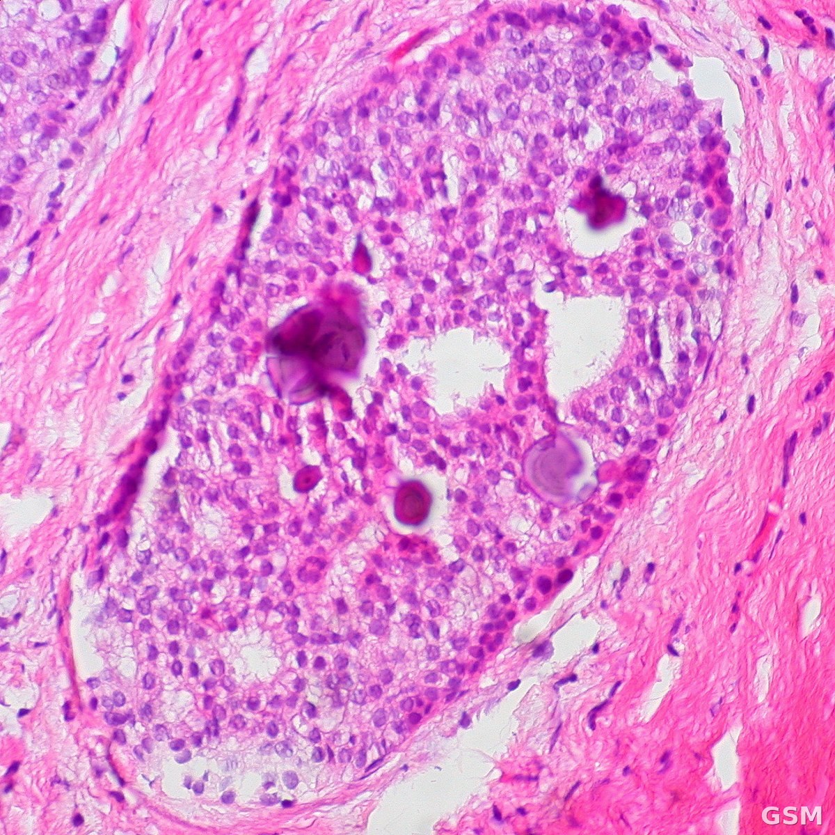
位于德克萨斯州哈里斯县休斯顿市的德克萨斯大学MD安德森癌症中心研究团队11月12日宣布,他们绘制了迄今最详细的导管原位癌(DCIS)周围细胞环境图谱之一,发现与浸润性乳腺癌相关的生物学特征出现的时间比此前认为的更早。研究当天发表于《Science》杂志。
DCIS是最常见的浸润前(0期)乳腺癌,异常细胞局限在乳腺导管内、尚未扩散至周围组织。随着乳腺X光等检查普及,检出的DCIS病例越来越多,但许多病变终生不会发展为浸润性癌,临床上一直难以判断哪些患者需要更积极的治疗、哪些患者可以安全地避免不必要的治疗。
这项研究由MD安德森系统生物学系主任 Nicholas Navin 博士、解剖病理学教授 Savitri Krishnamurthy 博士与贝勒医学院外科肿瘤学家 Alastair Thompson 博士共同领导,系统生物学博士后研究员 Siyuan He 博士与 Aatish Thennavan 博士为共同第一作者。团队运用先进的单细胞与空间分析技术,绘制了来自正常乳腺组织、DCIS和浸润性乳腺癌样本的逾85万个细胞,确认疾病进展过程中癌细胞及其周围微环境发生了广泛变化。研究还发现此前未知的「癌性间质环」:由支持组织与血管细胞构成的环状结构环绕DCIS病变,为肿瘤生长创造条件。
Navin 表示,DCIS被视为乳腺癌转为浸润性之前的一个截面,但并非所有DCIS都会进展为浸润性疾病,其病变周围的生物学环境一直难以考察;这份图谱揭示了可能帮助疾病转为浸润性的周围细胞与结构,未来有望帮助临床医生识别并针对性治疗浸润进展风险较高的DCIS患者,同时让低风险患者避免不必要的治疗。研究团队表示,仍需进一步研究验证这些发现,并评估其应用于未来诊断工具以及预防、治疗策略的潜力。
这项研究获得美国国家癌症研究所、德克萨斯州癌症预防与研究所(CPRIT)、Vivian L. Smith 基金会及MD安德森机构经费等支持。
(来源:德克萨斯大学MD安德森癌症中心)
MD Anderson Maps More Than 850,000 Cells Around Stage 0 Breast Cancer, Finding Invasive Features Emerge Earlier Than Thought
Researchers at The University of Texas MD Anderson Cancer Center in Houston, Harris County, Texas, announced on November 12 that they have created one of the most detailed maps to date of the cell environment surrounding ductal carcinoma in situ (DCIS), the most common form of preinvasive, or stage 0, breast cancer. The findings, published the same day in Science, suggest that biological features associated with invasive breast cancer begin to emerge earlier than previously known.
DCIS occurs when abnormal cells grow inside the breast milk ducts but have not yet spread into surrounding tissue. Mammograms and other breast examinations have increased the detection of DCIS cases, but many of these lesions never progress to invasive disease, making it challenging to determine which patients need more aggressive treatment and which may safely avoid unnecessary treatment.
The study was co-led by Nicholas Navin, Ph.D., chair of Systems Biology at MD Anderson; Savitri Krishnamurthy, M.D., professor of Anatomical Pathology; and Alastair Thompson, M.D., a surgical oncologist at Baylor College of Medicine, with Siyuan He, Ph.D., and Aatish Thennavan, Ph.D., postdoctoral fellows in Systems Biology, as co-first authors. Using advanced single-cell and spatial profiling technologies, the team mapped more than 850,000 cells from samples of normal breast tissue, DCIS and invasive breast cancer, identifying extensive changes in the cancer cells and the surrounding microenvironment as disease progressed. The study also uncovered previously unknown 'cancer stromal loops,' ring-like structures made of supportive tissue and vascular cells that surround DCIS lesions and create conditions that support tumor growth.
'DCIS is considered a snapshot before breast cancer becomes invasive, but not all DCIS cases progress to invasive disease, and the biology surrounding these lesions has been challenging to examine,' Navin said. 'This atlas reveals the surrounding cells and structures that may help disease become invasive. In the future, these findings may allow clinicians to better identify and treat patients with DCIS who are at higher risk of invasive progression while also avoiding unnecessary treatments for those at low risk.'
The researchers said additional research is needed to validate the findings and evaluate their potential use in future diagnostic tools and prevention or treatment strategies. The research was supported in part by the National Cancer Institute, the Cancer Prevention and Research Institute of Texas (CPRIT), the Vivian L. Smith Foundation and UT MD Anderson institutional funding.
(Source: The University of Texas MD Anderson Cancer Center)
来源:德克萨斯大学MD安德森癌症中心
发布:GSM·美国(发布链接)
图片来源:图:乳腺导管原位癌(DCIS)伴钙化的病理组织切片图像。照片:Ed Uthman,CC BY 2.0,经 Wikimedia Commons。 / Histology image of breast ductal carcinoma in situ (DCIS) with calcifications. Photo: Ed Uthman, CC BY 2.0, via Wikimedia Commons.
GSM Global · 本篇英文版 English edition of this story